
Welcome to the Katada Laboratory.


Established in September 2025 .
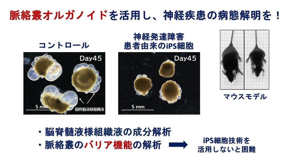
By utilizing iPS cell technology and mouse models, we aim to elucidate brain development, as well as the pathogenesis of neurological diseases, from the choroid plexus, a hidden tissue in the brain.
News
2026/06/08 ドイツ・ミュンヘン大学から学生を迎え、1か月間のインターンシップがスタートしました。
2026/05/31 iPS細胞20周年記念 公開シンポジウムで堅田が講演、山下がポスター発表させて頂きました。
2026/04/01 助教:山嵜絵海、特別研究学生:Kelren Rodrigues、研究生:楊玉婷が研究室に加わりました。
2026/03/14 第19回神経発生討論会@京都工業繊維大学で、Kelren Rodriguesが発表しました。
2026/01/01 研究員:山下りえが堅田研究室に加わりました。
2025/12/04 第48回日本分子生物学会年会「加齢に伴う器官変容のメカニズム」で、堅田が発表しました。
2025/11/04 第98回日本生化学会大会「細胞内外のオルガネラが織りなす多彩な脳内制御」で、堅田が発表しました。
2025/10/01 九州大学からの研究指導委託学生として、魚津孝允、有村崚が加わりました。
2025/09/27 中日バイオ医療・生命科学技術と産業融合フォーラムにて、堅田が発表しました。
2025/09/01 京都大学iPS細胞研究所 増殖分化機構研究部門に堅田研究室が発足しました。
The Hidden Tissue of the Brain:
Tackling Neurological Disorders through the Choroid Plexus
Research
Projects

Brain development
The cerebrospinal fluid environment created by the choroid plexus is a crucial signaling source supporting brain development during the embryonic stage. Our laboratory studies how trophic and growth factors secreted from the choroid plexus are received via the primary cilia of neural stem cells, and how they control cell fate and cerebral cortex formation. By utilizing single-cell multiohmics analysis, we aim to clarify how individual neural stem cells interpret information from the external environment and convert it into developmental programs, thereby gaining a new understanding of brain development.
Homeostasis
In adults, the choroid plexus plays a crucial role in maintaining homeostasis of the brain environment as the blood-CSF barrier. The blood vessels within the choroid plexus are composed of fenestrated vascular endothelium with high permeability, while choroid plexus epithelial cells strictly control the transport of substances into the brain. This barrier function not only supports the normal functioning of the brain but has also recently attracted attention as a drug delivery pathway to the brain. Our laboratory is recreating the human blood-CSF barrier using iPS cell-derived choroid plexus organoids and is conducting research on its barrier function, substance transport mechanisms, and potential applications in drug discovery.
Aging and neurological diseases
The choroid plexus is an essential tissue for brain development and homeostasis, and it has become clear that genetic changes and age-related functional decline in the choroid plexus have a significant impact on neurological function. Our laboratory is working to elucidate the molecular mechanisms by which abnormalities in the choroid plexus are involved in neurological diseases, using choroid plexus organoids created from patient-derived iPS cells and disease model mice. Furthermore, we aim to develop novel therapies for neurological disorders by targeting the choroid plexus and restoring or modulating its function.

Publications
Rodrigues K, Yamashita R, Katada S*
“The choroid plexus-cerebrospinal fluid axis as a lifespan regulator of neural stem cells and circuit plasticity”
Frontiers in Neural Circuits, 2026; 20:1818927 DOI:10.3389/fncir.2026.1818927
Katada S, Rodrigues K, Nakashima K*
“The influence of the choroid plexus on brain function: beyond its role in cerebrospinal fluid production”
Inflamm. Regen., 2025; 45(1):20 DOI:10.1186/s41232-025-00386-1
Nakagawa T, Hata K, Izumi Y, Nakashima H, Katada S, Matsuda T, Babba T, Nakashima K*
“E3 ubiquitin ligase RMND5A maintains the self-renewal state of human neural stem/precursor cells by regulating Wnt and mTOR signaling pathways”
FEBS Letters, 2025;599:10 DOI:10.1002/1873-3468.70067
Uotsu T, Nakashima K, Katada S*
“Multifaceted regulation of neural stem cell fate in the developing brain”
European Society of Medicine, 2024; 12: 1-17.
Uchihara Y, Sato H, Kawabata-Iwakawa R, Katada S, Gu W, Kakoti S, Yamauchi M,
Kato R, Gondhowiardjo S, Hosen N, Yasuhara T, & Shibata A*
“DNA damage promotes HLA class I presentation by stimulating a pioneer round of translation associated antigen production”
Mol Cell, 2022; 82: 2557-70. DOI: 10.1016/j.molcel.2022.04.030
Katada S*, Takouda J, Nakagawa T, Honda M, Igarashi K, Imamura T, Ohkawa Y,
Sato S, Kurumizaka H, & Nakashima K*
“Neural stem/precursor cells dynamically change their epigenetic landscape to differentially respond to BMP signaling for fate switching during brain development”
Genes & Development, 2021; 35: 1431-44. DOI:10.1101/gad.348797.121
Wittmann MT, Katada S, Sock E, Kirchner P, Ekici A, Wegner M, Nakashima K, Lie DC*, & Reis A*
“scRNA sequencing uncovers a TCF4-dependent transcription factor network regulating commissure development in mouse”
Development, 2021; 148: Dev196022. DOI: 10.1242/dev.196022.
Takouda J, Katada S*, Imamura T, Sanosaka T & Nakashima K*
“SoxE group transcription factor Sox8 promotes astrocytic differentiation of neural stem/precursor cells downstream of Nfia”
Pharmacol Res. & Perspectives, 2021; 9: e00749. DOI: 10.1002/prp.2.749.
Nguyen QAT, Hillis D, Katada S, Harris T, Pontrello C, Garland T & Haga-Yamanaka S*
“Coadaptation of the chemosensory system with voluntary exercise behavior in mice”
PLOS ONE, 2020; 15: e0241758. DOI: 10.1371/journal.pone.0241758.
Nakagawa T, Wada Y, Katada S*, & Kishi Y*
“Epigenetic regulation for acquiring glial identity by neural stem cells during cortical development”
Glia, 2020; 68: 1554-1567. DOI: 10.1002/glia.23818.
Kawamura Y, Katada S, Noguchi H, Yamamoto H, Sanosaka T, Iihara K, Nakashima K*
“Synergistic induction of astrocytic differentiation by factors secreted from meninges in the mouse developing brain”
FEBS Letters, 2017; 591: 3709-3720. DOI: 10.1002/1873-3468.12881.
Honda M, Nakashima K, & Katada S*
“PRMT1 regulates astrocytic differentiation of embryonic neural stem/precursor cells”
J Neurochem., 2017; 142: 901-907. DOI: 10.1111/jnc.14123.
Takouda J, Katada S, & Nakashima K*
“Emerging mechanisms underlying astrogenesis in the developing mammalian brain”
Pro Jpn Acad Ser B Phys Biol Sci., 2017; 93: 386-398. DOI: 10.1007/978-3-319-93485-3_5.
Aguilar-Arnal L, Katada S, Oroco-Solis R, & Sassone-Corsi P*
“NAD-SIRT1 control of H3K4 trimethylation through circadian deacetylation of MLL1”
Nat Struct Mol Biol., 2015; 22: 312-318. DOI: 10.1038/nsmb.2990.
Kaneko-Goto T#, Sato Y#, Katada S#, Kinameri E, Yoshihara S, Nishiyori A, Kimura M, Fujita H, Touhara K, Reed RR & Yoshihara Y* (# equally contributed)
“Goofy coordinates the acuity of olfactory signaling”
J Neurosci., 2013; 33: 12987-12996. DOI: 10.1523/JNEUROSCI.4948-12.2013.
Katada S, Imhof A, & Sassone-Corsi P*
“Connecting Threads: Epigenetic and Metabolism”
Cell, 2012; 148, 24-28. DOI: 10.1016/j.cell.2012.01.001.
Masri S, Zocchi L, Katada S, Mora E, & Sassone-Corsi P*
“The circadian clock transcriptional complex: metabolic feedback intersects with epigenetic control”
Anal NY Acad Sci., 2012; 1264, 103-109. DOI: 10.1111/j.1749-6632.2012.06649.x.
Katada S & Sassone-Corsi P*
“The histone methyltranferase MLL1 permit the oscillation of circadian gene expression”
Nat Struct Mol Biol., 2010; 17: 1414-1421. DOI: 10.1038/nsmb.1961.
Grimaldi B, Bellet MM, Katada S, Astarita G, Hirayama J, Amin RH, Granneman JG, Piomeli D, Leff T, & Sassone-Corsi P*
“Per2 controls lipid metabolism by direct regulation of PPARg”
Cell Metab., 2010; 12: 509-520. DOI: 10.1016/j.cmet.c010.10.005.
Kato A, Katada S, & Touhara K*
“Amino acids involved in conformational dynamics and G protein coupling of an odorant receptor: targeting gain-of-function mutation”
J Neurochem., 2008; 107: 1261-70. DOI: 10.1111/j.1471-4159.2008.05693.x.
Katada S, Hirokawa T, & Touhara K*
“Exploring the odorant binding site of a G-protein-coupled olfactory receptor”
Curr Comput Aided Drug Des., 2008; 4, 123-131. DOI: 10.2174/157340908784533247
Oka Y, Katada S, Omura M, Suwa M, Yoshihara Y, & Touhara K*
“Odorant receptor map in the mouse olfactory bulb: in vivo sensitivity and specificity of receptor-defined glomeruli”
Neuron, 2006; 52: 857-69. DOI: 10.1111/j.1471-4159.2008.05693.x.
Katada S, Hirokawa T, Oka Y, Suwa M, & Touhara K*
“Structural basis for a broad but selective ligand spectrum of a mouse olfactory receptor: mapping the odorant-binding site”
J Neurosci., 2005; 25: 1806-15. DOI: 10.1523/JNEUROSCI.4723-04.2005.
Katada S, Tanaka M, & Touhara K*
“Structural determinants for membrane trafficking and G protein selectivity of a mouse olfactory receptor”
J Neurochem., 2004; 90: 1454-63. DOI: 10.1111/j.1471-4159.2004.02619.x.
Katada S, Nakagawa T, Kataoka H, & Touhara K*
“Odorant response assays for a heterologously expressed olfactory receptor”
Biochem Biophys Res Commun. 2003; 305: 964-9. DOI: 10.1016/s0006-291x(03)00863-5.